Showing posts with label CV. Show all posts

Wednesday, August 22, 2012

Management of VF arrest

Passive rewarming

  • Temperature is very important during rewarming as temperature commonly overshoots normal. Warming the patient too quickly or allowing continued shivering causes dangerous electrolyte shifts, leading to potentially lethal arrhythmias. 
  • Controlled rewarming of 0.15° to 0.5° C per hour is recommended. 
  • To maintain tight temperature control throughout rewarming a neuromuscular blockade is usually employed.  
  • Careful fluid monitoring during rewarming is crucial because of the vasodilation that accompanies a body temperature rise. Volume replacement may be needed to prevent fluid deficit and hypotension.
  • Electrolytes shift out of the cells back into the serum during rewarming, so frequent electrolyte monitoring is needed during this phase to prevent critically elevated levels. Slow, controlled rewarming allows the kidneys to excrete excess potassium, preventing hyperkalemia. 
  • Hypoglycemia can occur during rewarming as the insulin resistance of earlier hypothermia phases diminishes. Glucose levels must be monitored frequently, with insulin titration and dextrose boluses used as needed to maintain the patient within ordered ranges.

References

  • http://www.americannursetoday.com/article.aspx?id=8014&fid=7986
  • http://ccforum.com/content/16/S2/A25/ 

Sunday, May 9, 2010

AMIs and thrombolysis

ECG changes indicating AMI

  • High probability of MI: persistent ST elevation of ≥ 1 mm in two contiguous limb leads or ST-segment elevation of ≥ 2 mm in two contiguous chest leads or the presence of new LBBB.
  • Intermediate probability of MI: are ST depression, T-wave inversion, and other nonspecific ST-T wave abnormalities.
  • Q waves = old MI

DDxes

Management options

  • Patients with persistent ST elevation should be considered for reperfusion therapy (thrombolysis or primary PCI).
  • Those without ST elevation will be diagnosed with either NSTEMI if cardiac marker levels are elevated or with unstable angina if serum cardiac marker levels provide no evidence of myocardial injury. Patients presenting with no ST-segment elevation are not candidates for immediate thrombolytics but should receive anti-ischemic therapy and may be candidates for PCI urgently or during admission.

Medical Management

  • Aspirin (300 mg) should be given unless already taken or contraindicated (grade A recommendation), and should preferably be given early (eg, by emergency or ambulance personnel).
  • Clopidogrel should be given in addition to aspirin for patients undergoing PCI with a stent (loading-dose of 300–600 mg), or for fibrinolytic therapy (300 mg). Clopidogrel 75 mg daily should be continued for at least a month after fibrinolytic therapy, and for up to 12 months after stent implantation, depending on the type of stent.
  • Antithrombin therapy to inhibit the coagulation cascade, and for patients underdoing PCI. For patients getting streptokinase, whether to heparinise depends on the anti-thrombotic agent. Clexane (enoxaparin) bolus should be dosed at 0.75 mg/kg.
  • Administer a platelet glycoprotein (GP) IIb/IIIa-receptor antagonist (eptifibatide, tirofiban, or abciximab) in addition to aspirin and unfractionated heparin, to patients with continuing ischemia or with other high-risk features and to patients in whom PCI is planned.
  • An ACE inhibitor (Captopril) should be given orally within the first 24 hours of STEMI to patients with anterior infarction, pulmonary congestion, or left ventricular ejection fraction (LVEF) less than 40% in the absence of hypotension.
  • An angiotensin receptor blocker (valsartan or candesartan) should be administered to patients with STEMI who are intolerant of ACE inhibitors and who have either clinical or radiological signs of heart failure or LVEF less than 40%.

Contraindications for fibrinolytic use in STEMI

Absolute contraindications:
  • Prior intracranial hemorrhage (ICH)
  • Known structural cerebral vascular lesion
  • Known malignant intracranial neoplasm
  • Ischemic stroke within 3 months
  • Suspected aortic dissection
  • Active bleeding or bleeding diathesis (excluding menses)
  • Significant closed-head trauma or facial trauma within 3 months
Relative contraindications:
  • History of chronic, severe, poorly controlled hypertension
  • Severe uncontrolled hypertension on presentation (SBP >180 mm Hg or DBP >110 mm Hg)
  • Traumatic or prolonged (>10 min) CPR or major surgery less than 3 weeks
  • Recent (within 2-4 wk) internal bleeding
  • Noncompressible vascular punctures
  • For streptokinase/anistreplase - prior exposure or prior allergic reaction to these agents
  • Pregnancy
  • Active peptic ulcer
  • Current use of anticoagulant (eg, warfarin sodium) that has produced an elevated international normalized ratio (INR) >1.7 or prothrombin time (PT) >15 seconds

Follow-up Patient Care

  • Patients should continue to receive beta-blockers, nitrates, and heparin, as indicated.
  • ACE inhibitors have been shown to improve survival rates in patients who have experienced an MI. In the acute setting, afterload reduction from ACE inhibitors may reduce the risk of CHF and sudden death.

References

Tuesday, January 12, 2010

CHAD2 (CHADS) score

The CHADS score is a clinical prediction rule for estimating the risk of stroke in patients with non-rheumatic atrial fibrillation (AF) and is used to determine the degree of anticoagulation therapy required.

To Score...

C ongestive heart failure (1 point)
H ypertension > 160mmHg systolic (or treated hypertension) (1 point)
A ge > 75 (1 point)
D iabetes (1 point)
S - previous stroke or TIA (2 points)

Risk of stroke based on CHADS score

Recommendations for anticoagulation

  • High risk (score >= 2) - warfarin (unless contrainidcated)
  • Moderate risk (score 1) - aspirin or warfarin
  • Low risk (score 0) - aspirin

References

  • http://en.wikipedia.org/wiki/CHADS_Score
  • http://www.cardiology.org/tools/risk_of_stroke_AF.html

Tuesday, August 28, 2007

Microalbumuria

Screening for microalbumuria in non-diabetics is a good way to screen for CV risks. The microalbumuria is caused by endothelial dysfunction.

SAAB drugs for use in acute coronary syndrome

  • Statin
  • Ace inhibitor
  • Aspirin
  • Beta blocker

Treatment of endocarditis

Endocarditis requires 6 weeks of IV antibiotic treatment. This length of time is required because it is difficult for antibiotics to penetrate to the valve cusps which are relatively avascular.

Thursday, August 23, 2007

Calcium channel blockers

Mechanism of action

Calcium channel blockers have a negative inotropic effect - they decrease the force of contraction of the myocardium.

They block L-type voltage gated calcium channels in the heart and blood vessels. This prevents calcium levels from increasing as much in the cells when stimulated, leading to less contraction.

They also decrease total peripheral resistance by dilating the blood vessels, and decreasing cardiac output by lowering the force of contraction. Because resistance and output drop, so does blood pressure. With low blood pressure, the heart does not have to work as hard; this can ease problems with cardiomyopathy and coronary disease.

Unlike with beta-blockers, the heart is still responsive to sympathetic nervous system stimulation, so blood pressure can be maintained more effectively.

Many calcium channel blockers also slow down the conduction of electrical activity within the heart by blocking the calcium channel during the plateau phase of the action potential of the heart. This causes a lowering of the heart rate and may cause heart blocks (negative chronotropic effect) of calcium channel blockers.

Classes

There are 2 classes of CCBs:
  1. Dihydropyridines

    • Used to reduce systemic vascular resistance and arterial pressure, but are not used to treat angina because the vasodilation and hypotension can lead to reflex tachycardia.
    • This CCB class is easily identified by the suffix "-pine" e.g. Amlodepine, Felodipine.

  2. Non-dihydropyridines

    • Relatively selective for myocardium, reduce myocardial oxygen demand and reverse coronary vasospasm, and are often used to treat angina.
    • They have minimal vasodilatory effects compared with dihydropyridines.
    • Action is intracellular.
    • E.g. verapamil.

Indications

  • Atrial fibrillation or flutter - to control heart rate via negative chronotropic effect.

Contraindications

  • Avoided (or used with caution) in individuals with cardiomyopathy due to negative inotropic effect.
  • Non-dihydropyridine CCBs should not be combined with beta-blockers because they are both negative inotropes and affect the AV node.
References:
  • Wikpedia, http://en.wikipedia.org/wiki/Calcium_channel_blocker

Sunday, July 22, 2007

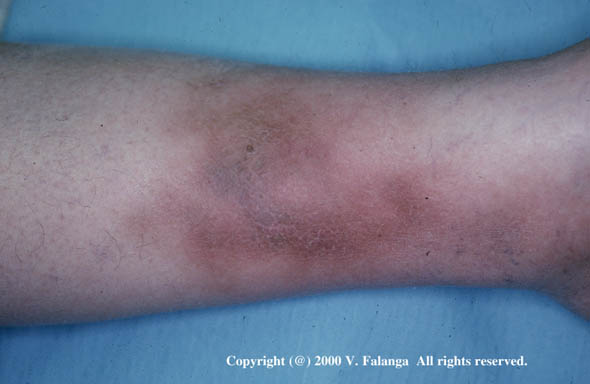
Lipodermatosclerosis (LDS)

  • LDS literally means "scarring of the skin and fat" and is a slow process that occurs over a number of years.
  • Occurs in patients with long-standing venous disease resulting in chronic venous insufficiency.
  • Affects the skin just above the ankle, usually on the inside surface.
  • Over time the skin becomes brown, smooth, tight and often painful.
  • The precise mechanism of LDS is not fully understood, but we do know that it is caused by an excessively high venous pressure in the subcutaneous veins in the lower leg.
References:
  • http://www.simondodds.com/Venous/LDS.htm
  • http://www.bu.edu/woundbiotech/wounds/UncommonWounds%20Gallery/pages/19..htm

Chronic Venous Insufficiency (CVI)

Symptoms

  • Varicose veins
  • Ulceration or skin breakdown
  • Lipodermatosclerosis
  • Reddened or discolored skin on the leg
  • Oedema

Risk factors

  • Heredity
  • Obesity
  • Pregnancy
  • Sedentary lifestyle
  • Smoking
  • Jobs requiring long periods of standing or sitting in one place
  • Age and sex (women in their 50s are more prone to developing CVI)
  • Incompetent valves
  • previous DVT

Pathophysiology

  • Increased venous pressure transcends the venules to the capillaries, impeding flow.

  • Low-flow states within the capillaries cause leukocyte trapping.

  • Trapped leukocytes release proteolytic enzymes and oxygen free radicals, which damage capillary basement membranes.

  • Plasma proteins,such as fibrinogen, leak into the surrounding tissues, forming a fibrin cuff.

  • Interstitial fibrin and resultant oedema decrease oxygen delivery to the tissues, resulting in local hypoxia.

  • Inflammation and tissue loss result. Ulceration may occur.

Management

  • Leg elevation
  • Elastic compression therapy - especially with older patients you need to ensure they are actually able to put the compression stockings on for themselves.
  • Sclerotherapy - chemically scarring the veins from the inside out so that they can then no longer fill with blood. Blood that would normally return to the heart through these veins returns to the heart through others. The body eventually absorbs the veins that received the injection.
  • Vein stripping
  • Deep vein surgery - note that surgical treatment is reserved for those with discomfort or ulcers refractory to medical management.
  • Valve repair

Saturday, July 14, 2007

Ankle-Brachial Index

The Ankle – Brachial Index (ABI) is a ratio of the systolic blood pressure measured simultaneously in the leg and arm. This test is done to screen for peripheral arterial disease (PAD) of the legs.

What the values mean:
  • An ABI ratio less than 0.8 implies significant arterial obstruction.
  • A ratio of 0.5 or less implies critical obstruction.
  • A ratio of < 0.3 implies impending gangrene.
Patients with PAD should not wear compression stockings due to high risk of ischaemia.

References:

Management of adult cardiorespiratory arresst

Wednesday, July 4, 2007

Giant cell arteritis (GCA)

Giant cell arteritis is a vasculitis of large and medium size vessels. Although it can affect arteries in the neck, upper body and arms, it occurs most often in the arteries in the temples. For this reason, giant cell arteritis is sometimes called temporal arteritis or cranial arteritis.

Giant cell arteritis is also known as granulomatous arteritis — a reference to a particular type of inflammation it causes.

Epidemiology & Aetiology

  • Adults older than age 50 are at greatest risk of giant cell arteritis.
  • Women and caucasians are most commonly affected.
  • The exact cause isn't known, but researchers believe that genetic, viral and environmental factors may play roles in the inflammation.

Clinical presentation

Giant cell arteritis frequently causes headaches, jaw pain, and blurred or double vision, but the most serious potential complications are blindness and, less often, stroke. These problems occur when swelling in the arteries impairs blood flow to the eyes or brain.

The onset of the symptoms tends to be gradual and includes low grade fever, fatigue, weakness and weight loss.

  • A new headache, mild or severe, occurs in at least two-thirds of patients with the pain tending to be located over the sides of the head in front of the ears but may be frontal or other located.
  • Nearly one-half of patients suffer from jaw claudication after chewing.
  • Impaired vision is often an early manifestation of the disease.
  • Permanent partial or complete loss of vision in one or both eyes has been observed in 15-20 % of patients. It is rare for patients to become completely blind in both eyes.
  • Polymyalgia rheumatica, which is characterized by pain in the shoulders and hips, is closely linked to GCA, occurring in about 40-50 % of patients.

Investigations

  • ESR – elevated in most patients with GCA.
  • Temporal artery biopsy
  • Suggested in all cases of suspected GCA even if the diagnosis may appear "classic".
  • The biopsy is of low risk, causes very little pain, and often leaves little or no scar.
  • After the use of a topical numbing medication (the same one used by a dentist), a small part of the temporal artery from under the scalp is removed.
  • Other ways to diagnose GCA include: ultrasonography, angiographic examination, CT scanning and MR angiography, high resolution MRI and position emission tomography (PET).

Management

  • Although there's no cure for giant cell arteritis, immediate treatment with corticosteroid medications usually relieves symptoms and prevents loss of vision.
  • Glucocorticoid treatment should be instituted immediately once the diagnosis of GCA is established.
  • Daily dosing is more effective than alternate day dosing. The response usually occurs within two to four weeks after the institution of therapy.
  • The diagnosis should be reevaluated in patients who are resistant to adequate steroid therapy.
  • Steroid withdrawal can begin once clinical remission has been induced.
  • Relapses are seen more frequently in the first year or two of the disease.
  • Relapses often necessitate increased dosage or prolonged steroid treatment. Some researchers have suggested that the addition of methotrexate may be steroid-sparing while others have not demonstrated any benefit. However the routine addition of methotrexate to glucocorticoid therapy for GCA is not recommended. The efficacy of other cytotoxic drugs, dapsone, antimalarials, etanercept, and penicillamine has not been studied adequately although they have been reported to be helpful in some case reports.
  • The finding of an increased risk of visual loss in patients with GCA and thrombocytosis (increase of the number of platelets in the blood), has led some to suggest the addition of drugs like aspirin for patients with high platelet counts, but there is not a lot of data to prove that this may reduce brain/skull problems.

References:
  • “Giant cell arteritis”, mayoclinic.com, http://www.mayoclinic.com/health/giant-cell-arteritis/DS00440
  • “Giant Cell Arteritis (Temporal Arteritis)”, Vasculitis Foundation, http://www.vasculitisfoundation.org/giantcellarteritis

Relationship between pulmonary embolism (PE) and atrial fibrillation (AF)

If PE and AF occur together it is most likely that the PE is the cause of the AF.

The mechanism of the AF is said to be acute right ventricular dilatation with "strain" due to the embolus in the pulmonary circulation creating backpressure into the right ventricle.

If a patient presents with unexplained AF, look for an accompanying PE as the cause.

It is postulated that AF can cause PE if a clot originates in the right atrium rather than the left, but this is less common than clots originating from the right ventricle and more studies need to be done in this area.

References:
  • Flegel K., When atrial fibrillation occurs with pulmonary embolism, is it the chicken or the egg?, CMAJ 1999;160:1181-2


Monday, April 30, 2007

Stigmata of infective endocarditis

  • Fever (often spiking)
  • Continuous presence of micro-organisms in the bloodstream in serial collection of blood cultures
  • Vegetations on valves on echocardiography, which sometimes can cause a new or changing heart murmur, particularly murmurs suggestive of valvular regurgitation
  • Vascular phenomena:
  • Septic emboli - mitral regurg -> LA dilatation -> ectopic foci -> stasis in LA -> formation of thrombi -> circulatory problems such as stroke or gangrene of fingers
  • Janeway lesions (painless hemorrhagic cutaneous lesions on the palms and soles)
  • hemorrhage: intracranial hemorrhage, conjunctival hemorrhage, splinter hemorrhages
  • Immunologic phenomena: Glomerulonephritis, Osler's nodes (painful subcutaneous lesions in the distal fingers), Roth's spots on the retina, positive serum rheumatoid factor


References:

  • http://en.wikipedia.org/wiki/Endocarditis

Tuesday, April 24, 2007

Management of AF


References:
  • http://www.aafp.org/afp/20020715/249.html

Monday, April 23, 2007

S1Q3T3

What it is

The S1Q3T3 is the ECG manifestation of acute pressure and volume overload of the right ventricle.

It is characterised by:
  • Lead I - an S wave signifying a complete or more often incomplete RBBB.
  • Lead III - a Q wave, slight ST elevation, and an inverted T wave. These findings are due to the pressure and volume overload over the right ventricle which causes repolarization abnormalities.

Causes

Any cause of acute cor pulmonale can cause the S1Q3T3 finding on the ECG. This includes PE, acute bronchospasm, pneumothorax, and other acute lung disorders. In addition, transient LPFB may cause this finding as well.

What this means for Dx'ing PE

The ECG is often abnormal in PE, but findings are not sensitive & not specific. S1Q3T3 pattern is present in only in 20% of cases of PE.

The ECG is a poor diagnostic tool for PE. The greatest utility of the ECG in the patient with suspected PE is ruling out other potential life-threatening diagnoses such as MI.


References:
  • http://medicine.ucsf.edu/housestaff/Chiefs_cover_sheets/ecg_pe.pdf
  • http://www.ispub.com/ostia/index.php?xmlFilePath=journals/ijem/vol3n1/cor.xml

Monday, February 26, 2007

Arterial vs venous clots

Arterial clots

Arteries are thick blood vessels with fast flowing blood. Blood clots in arteries are typically triggered by underlying arteriosclerosis (roughening of the artery wall). Blood platelets get stuck to the roughened blood vessel wall and form a clot. Thus, the medication of choice in trying to prevent thrombosis in arteries are medications that act against platelets. The following medications are anti-platelet drugs:
  • Aspirin (= ASA)
  • Plavix (= Clopidogrel)
  • Ticlid (= Ticlopidine)
  • Aggrenox (= aspirin plus dipyridamole)
By interfering with platelet function, these drugs increase the patient's risk of bleeding, even though to a lesser degree than coumadin. The INR is not influenced by these drugs and vitamin K intake does not influence their effect.

Venous clots

Veins are thin blood vessels with slow flowing blood. Blood clots that form in veins (DVT, pulmonary embolism) are mainly made up of clotting proteins; platelets do not play a big role in venous clots. Warfarin is an effective anticoagulant by preventing the production of clotting factors in the liver, increasing the INR. It is therefore the drug of choice in venous thrombosis. Anti-platelet drugs do not play much of a role in preventing venous clots.

Occasionally, clots in arteries originate from one of the two left heart chambers and travel from there with the blood stream to the brain, the retina, or the extremities. This typically happens in atrial fibrillation. Such a clot is an arterial embolism that resembles the type of clots seen in veins i.e. they have little platelet participation. They are therefore best treated with warfarin, not with anti-platelet drugs, even though they are clots in arteries.

References: