Friday, August 24, 2012

Dead Space Ventilation

Dead space is the volume of air which is inhaled but does not take part in gas exchange either because it remains in the conducting airways or in alveoli that are poorly perfused i.e. not all the air in each breath is able to be used for the exchange of oxygen and carbon dioxide.

 The total dead space (also known as physiological dead space) is the sum of the anatomical dead space plus the alveolar dead space.

Anatomic dead space

Approx one-third tidal volume ~150mL in a healthy adult.

Alveolar dead space

  • Sum of the volumes of alveoli which have little or no blood flowing through their adjacent pulmonary capillaries i.e., alveoli that are ventilated but not perfused, and where, as a result, no gas exchange can occur.
  • Alveolar dead space is negligible in healthy individuals, but can increase dramatically in some lung diseases due to ventilation-perfusion mismatch.


References


  • http://en.wikipedia.org/wiki/Dead_space_(physiology)
  • http://www.paramedicine.com/pmc/End_Tidal_CO2.html

Plateau effect

Takes 4-5 half lifes for a medication to reach plateau effect.

Wednesday, August 22, 2012

Cryoprecipitate

  • A frozen blood product prepared from plasma
  • Each 15 mL unit typically contains 100 IU of factor VIII, 250 mg of fibrinogen, as well as von Willebrand factor (vWF) and factor XIII.
  • Used commonly for DIC to keep fibrinogen levels > 1.0.
  • 1 unit of cryo per 5kg patient weight will increase fibrinogen by about 100 mg/dL. Therefore number of bags = 0.2 x weight (kg) to provide about 100mg/dL fibrinogen.
  • Many institutions use a standard dose of 10 units and then repeat if needed.

References

  • http://en.wikipedia.org/wiki/Cryoprecipitate
  • http://reference.medscape.com/drug/cryo-cryoprecipitate-999498
  • http://www.transfusion.com.au/sites/default/files/iTRANSFUSE%202.2%20CRYO.pdf
  • http://www.perthhaematology.com.au/cryo.ht

Anuric renal failure

Anuric renal failure


  • Urea rises by 10 per day, Cr rises by 100 per day

References

  • http://www.rph.wa.gov.au/nephrology/Acute_renal_failure.html
  • http://www.doctorslounge.com/nephrology/diseases/acute_renal_failure.htm
  • http://web.up.ac.za/sitefiles/file/45/1335/4101/Tuesday%20Academic%20Meetings/N%20Grabowski%204%20May%202011%20diagnosis%20of%20renal%20failure.pdf

Management of VF arrest

Passive rewarming

  • Temperature is very important during rewarming as temperature commonly overshoots normal. Warming the patient too quickly or allowing continued shivering causes dangerous electrolyte shifts, leading to potentially lethal arrhythmias
  • Controlled rewarming of 0.15° to 0.5° C per hour is recommended. 
  • To maintain tight temperature control throughout rewarming a neuromuscular blockade is usually employed.  
  • Careful fluid monitoring during rewarming is crucial because of the vasodilation that accompanies a body temperature rise. Volume replacement may be needed to prevent fluid deficit and hypotension.
  • Electrolytes shift out of the cells back into the serum during rewarming, so frequent electrolyte monitoring is needed during this phase to prevent critically elevated levels. Slow, controlled rewarming allows the kidneys to excrete excess potassium, preventing hyperkalemia
  • Hypoglycemia can occur during rewarming as the insulin resistance of earlier hypothermia phases diminishes. Glucose levels must be monitored frequently, with insulin titration and dextrose boluses used as needed to maintain the patient within ordered ranges.

References

  • http://www.americannursetoday.com/article.aspx?id=8014&fid=7986
  • http://ccforum.com/content/16/S2/A25/ 

Wednesday, May 4, 2011

DCR

DCR = cardioversion (direct current reversion)

References

Pantoprazole infusion

For actively bleeding ulcers give pantoprazole bolus followed by infusion.
  • LOading dose: Pantoprazole 80mg IV in 100ml of NaCl 0.9% or 5% glucose over 20 - 30 minutes
  • Infusion:
    • Pantoprazole 200mg in Dextrose 5% 500mL at 20mL/hr (each mL is 0.4mg , 20mL/hr = 8mg/hr, 1mg =2.5mL)
    • Pantoprazole 80mg in N/Saline 100mL at 10mL/hr (each mL is 0.8mg , 10mL/hr = 8mg/hr,1mg=1.25mL)

References

Tuesday, January 25, 2011

Blood Gas normal values


ArterialVenous
pH7.35-7.457.3-7.35
pCO235-4545-46 (good representation of ventilation)
pO280-10020-80 (uninterprable)
SaO295-100
HCO3-22-28
BE+/- 3


Tuesday, December 7, 2010

Resources for lumbar puncture

  • http://www.med.uottawa.ca/procedures/lp/index.htm
  • http://www.articlealley.com/article_596234_17.html
  • http://www.unboundmedicine.com/harrisons/ub/view/Harrisons-Manual-of-Medicine/148408/all/Lumbar_Puncture,

Tuesday, November 30, 2010

Resources for central line insertion

Central lines in general

  • http://egret.psychol.cam.ac.uk/medicine/Central_line_insertion.pdf
  • http://www.nda.ox.ac.uk/wfsa/html/u12/u1213_01.htm
  • http://www.proceduresconsult.com/medical-procedures/central-venous-line-placement-AN-procedure.aspx

Internal jugular lines

  • http://www.anwresidency.com/simulation/guide/ij.html

Sunday, May 9, 2010

AMIs and thrombolysis

ECG changes indicating AMI

  • High probability of MI: persistent ST elevation of ≥ 1 mm in two contiguous limb leads or ST-segment elevation of ≥ 2 mm in two contiguous chest leads or the presence of new LBBB.
  • Intermediate probability of MI: are ST depression, T-wave inversion, and other nonspecific ST-T wave abnormalities.
  • Q waves = old MI

DDxes

Management options

  • Patients with persistent ST elevation should be considered for reperfusion therapy (thrombolysis or primary PCI).
  • Those without ST elevation will be diagnosed with either NSTEMI if cardiac marker levels are elevated or with unstable angina if serum cardiac marker levels provide no evidence of myocardial injury. Patients presenting with no ST-segment elevation are not candidates for immediate thrombolytics but should receive anti-ischemic therapy and may be candidates for PCI urgently or during admission.

Medical Management

  • Aspirin (300 mg) should be given unless already taken or contraindicated (grade A recommendation), and should preferably be given early (eg, by emergency or ambulance personnel).
  • Clopidogrel should be given in addition to aspirin for patients undergoing PCI with a stent (loading-dose of 300600 mg), or for fibrinolytic therapy (300 mg). Clopidogrel 75 mg daily should be continued for at least a month after fibrinolytic therapy, and for up to 12 months after stent implantation, depending on the type of stent.
  • Antithrombin therapy to inhibit the coagulation cascade, and for patients underdoing PCI. For patients getting streptokinase, whether to heparinise depends on the anti-thrombotic agent. Clexane (enoxaparin) bolus should be dosed at 0.75 mg/kg.
  • Administer a platelet glycoprotein (GP) IIb/IIIa-receptor antagonist (eptifibatide, tirofiban, or abciximab) in addition to aspirin and unfractionated heparin, to patients with continuing ischemia or with other high-risk features and to patients in whom PCI is planned.
  • An ACE inhibitor (Captopril) should be given orally within the first 24 hours of STEMI to patients with anterior infarction, pulmonary congestion, or left ventricular ejection fraction (LVEF) less than 40% in the absence of hypotension.
  • An angiotensin receptor blocker (valsartan or candesartan) should be administered to patients with STEMI who are intolerant of ACE inhibitors and who have either clinical or radiological signs of heart failure or LVEF less than 40%.

Contraindications for fibrinolytic use in STEMI

Absolute contraindications:
  • Prior intracranial hemorrhage (ICH)
  • Known structural cerebral vascular lesion
  • Known malignant intracranial neoplasm
  • Ischemic stroke within 3 months
  • Suspected aortic dissection
  • Active bleeding or bleeding diathesis (excluding menses)
  • Significant closed-head trauma or facial trauma within 3 months
Relative contraindications:
  • History of chronic, severe, poorly controlled hypertension
  • Severe uncontrolled hypertension on presentation (SBP >180 mm Hg or DBP >110 mm Hg)
  • Traumatic or prolonged (>10 min) CPR or major surgery less than 3 weeks
  • Recent (within 2-4 wk) internal bleeding
  • Noncompressible vascular punctures
  • For streptokinase/anistreplase - prior exposure or prior allergic reaction to these agents
  • Pregnancy
  • Active peptic ulcer
  • Current use of anticoagulant (eg, warfarin sodium) that has produced an elevated international normalized ratio (INR) >1.7 or prothrombin time (PT) >15 seconds

Follow-up Patient Care

  • Patients should continue to receive beta-blockers, nitrates, and heparin, as indicated.
  • ACE inhibitors have been shown to improve survival rates in patients who have experienced an MI. In the acute setting, afterload reduction from ACE inhibitors may reduce the risk of CHF and sudden death.

References

Friday, March 12, 2010

Guardianship

If a patient refuses treatment...
  • If NOK agrees not to treat → OK but document it
  • If NOK thinks treatment should occur → need to go to guardianship tribunal

Sunday, January 17, 2010

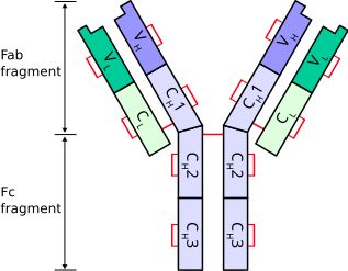
Antibodies

Antibodies consist of two Ig heavy chains (blue) linked by disulfide bonds to two Ig light chains (green).


Heavy chains

Heavy chains define the class of immunoglobulin. There are 5 types of heavy chains:
  1. α (Ig A)
  2. δ (Ig D)
  3. ε (Ig E)
  4. γ (Ig G)
  5. μ (Ig M)
The immunoglobulin heavy chain gene complex has been assigned to chromosome 14.

Light chains

There are 2 types of light chains:
  1. Lambda (λ) - encoded by a gene on chromosome 22
  2. Kappa (κ) - encoded by a gene on chromosome 2
Ig light chains produced in neoplastic plasma cells (e.g. in multiple myeloma) are called Bence Jones proteins.

References

  • http://en.wikipedia.org/wiki/Immunoglobulin_heavy_chain
  • http://en.wikipedia.org/wiki/Immunoglobulin_light_chain
  • http://en.wikipedia.org/wiki/Multiple_myeloma#Pathophysiology
  • http://www3.interscience.wiley.com/journal/120047597/abstract?CRETRY=1&SRETRY=0

Friday, January 15, 2010

Well’s Criteria for DVT

The Score

  1. Active cancer (treatment within last 6 months or palliative) -- 1 point
  2. Calf swelling >3 cm compared to other calf (measured 10 cm below tibial tuberosity) -- 1 point
  3. Collateral superficial veins (non-varicose) -- 1 point
  4. Pitting edema (confined to symptomatic leg) -- 1 point
  5. Swelling of entire leg - 1 point
  6. Localized pain along distribution of deep venous system—1 point
  7. Paralysis, paresis, or recent cast immobilization of lower extremities—1 point
  8. Recently bedridden > 3 days, or major surgery requiring regional or general anesthetic in past 4 weeks—1 point
  9. Alternative diagnosis at least as likely—Subtract 2 points
Possible score -2 to 8

Interpretation

Score of 2 or higher - deep vein thrombosis is likely. Consider imaging the leg veins.
Score of less than 2 - deep vein thrombosis is unlikely. Consider blood test such as d-dimer test to further rule out deep vein thrombosis.

References

  • http://en.wikipedia.org/wiki/Deep_vein_thrombosis
  • http://emedicine.medscape.com/article/758140-overview

Tuesday, January 12, 2010

CHAD2 (CHADS) score

The CHADS score is a clinical prediction rule for estimating the risk of stroke in patients with non-rheumatic atrial fibrillation (AF) and is used to determine the degree of anticoagulation therapy required.

To Score...

C ongestive heart failure (1 point)
H ypertension > 160mmHg systolic (or treated hypertension) (1 point)
A ge > 75 (1 point)
D iabetes (1 point)
S - previous stroke or TIA (2 points)

Risk of stroke based on CHADS score

Recommendations for anticoagulation

  • High risk (score >= 2) - warfarin (unless contrainidcated)
  • Moderate risk (score 1) - aspirin or warfarin
  • Low risk (score 0) - aspirin

References

  • http://en.wikipedia.org/wiki/CHADS_Score
  • http://www.cardiology.org/tools/risk_of_stroke_AF.html

Friday, January 8, 2010

ABCD^2 (ABCD squared) post TIA stroke risk assessment tool

The Tool

AGE: greater than or equal to 60 years – 1 point
Blood Pressure: Systolic >= 140, diastolic >= 90 (when first assessed after TIA)1 point
Clinical Features: unilateral weakness2 points, isolated speech disturbance1 point, other – zero
Duration of TIA symptoms: greater than or equal to 60 minutes – 2 points, 10 to 59 minutes – 1 point, <10 minutes zero
Diabetes present – 1 point

Estimated two day stroke risks determined by the ABCD^2 score:

  • Score 6 to 7: High two day stroke risk (8.1%)
  • Score 4 to 5: Moderate two day stroke risk (4.1%)
  • Score 0 to 3: Low two day stroke risk (1.0%)
  • Score < 1: Very low two day stroke risk (0.0%)

In versus outapatient management

People with a high risk of stroke (ABCD2 score of 4 or above) should have:
  • Aspirin (300 mg daily) started immediately
  • specialist assessment and investigation within 24 hours of onset of symptoms
  • measures for secondary prevention introduced as soon as the diagnosis is confirmed, including discussion of individual risk factors
People who are at lower risk of stroke (ABCD2 score of 3 or below) should have:
  • Aspirin (300 mg daily) started immediately
  • specialist assessment and investigation as soon as possible, but definitely within 1 week of onset of symptoms
  • measures for secondary prevention introduced as soon as the diagnosis is confirmed, including discussion of individual risk factors

Investigations

  1. CT scan without enhancement should be done in all patients to exclude other causes of neurological deficit (e.g. hemorrhage, subdural hematoma)
    • The presence of an infarct on CT is highly predictive of subsequent stroke
    • Early CT showing hemorrhage makes carotid imaging unnecessary
  2. Carotid imaging should be done for all patients with symptoms in anterior circulation territory.
    • The presence of carotid disease is highly predictive of recurrent stroke.
    • Consider CT angiogram if Carotid Ultrasound cannot be obtained in reasonable time.
  3. ECG and occasionally Holter monitoring to detect atrial fibrillation.
  4. ECHO cardiogram for persons with suspect underlying cardiac abnormalities.
  5. Blood sugar to detect extremes in glucose levels.

References

  • http://bmhgt.com/2009/03/stroke-information-abcd2/
  • http://www.gpnotebook.co.uk/simplepage.cfm?ID=x20080723164438749131
  • http://www.palmedpage.com/Text_files/Neurology/ABCD/TIA%20Management.html
  • http://www.stroke.org/site/DocServer/NSA_ABCD2_tool.pdf?docID=5981

Thursday, December 17, 2009

Paediatric elbow trauma

You can tell the approximate age of a child from the degree of bone replacement of cartilage on x-ray. Approximately :
  • Capitellum 'appears' at 2
  • Radial head at 4
  • Medial condyle at 6
  • Trochlea at 8
  • Olecranon at 10
  • Lateral condyle at 12 years of age
The acronym CRMTOL is used to describe the usual order of appearance of all 6 elbow centers: capitellum, radial head, medial epicondyle, trochlea, olecranon, and lateral epicondyle.

References

Wednesday, November 18, 2009

Sedation tips

  • Don't use midazolam and olanzapine together as it can cause rapid onset respiratory depression. Clonazepam and olanzipine is a better combination.
  • Chlorpromazine is also good. Non-addictive, and comes as a liquid.

Wednesday, November 4, 2009

Blood Products timing

RBCs 1 unit q4h
FFP 1 unit q1h
Platelets 1 unit q30minutes

Thursday, September 17, 2009