Sunday, January 17, 2010

Antibodies

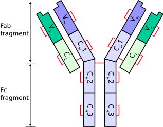
Antibodies consist of two Ig heavy chains (blue) linked by disulfide bonds to two Ig light chains (green).


Heavy chains

Heavy chains define the class of immunoglobulin. There are 5 types of heavy chains:
  1. α (Ig A)
  2. δ (Ig D)
  3. ε (Ig E)
  4. γ (Ig G)
  5. μ (Ig M)
The immunoglobulin heavy chain gene complex has been assigned to chromosome 14.

Light chains

There are 2 types of light chains:
  1. Lambda (λ) - encoded by a gene on chromosome 22
  2. Kappa (κ) - encoded by a gene on chromosome 2
Ig light chains produced in neoplastic plasma cells (e.g. in multiple myeloma) are called Bence Jones proteins.

References

  • http://en.wikipedia.org/wiki/Immunoglobulin_heavy_chain
  • http://en.wikipedia.org/wiki/Immunoglobulin_light_chain
  • http://en.wikipedia.org/wiki/Multiple_myeloma#Pathophysiology
  • http://www3.interscience.wiley.com/journal/120047597/abstract?CRETRY=1&SRETRY=0

Friday, January 15, 2010

Well’s Criteria for DVT

The Score

  1. Active cancer (treatment within last 6 months or palliative) -- 1 point
  2. Calf swelling >3 cm compared to other calf (measured 10 cm below tibial tuberosity) -- 1 point
  3. Collateral superficial veins (non-varicose) -- 1 point
  4. Pitting edema (confined to symptomatic leg) -- 1 point
  5. Swelling of entire leg - 1 point
  6. Localized pain along distribution of deep venous system—1 point
  7. Paralysis, paresis, or recent cast immobilization of lower extremities—1 point
  8. Recently bedridden > 3 days, or major surgery requiring regional or general anesthetic in past 4 weeks—1 point
  9. Alternative diagnosis at least as likely—Subtract 2 points
Possible score -2 to 8

Interpretation

Score of 2 or higher - deep vein thrombosis is likely. Consider imaging the leg veins.
Score of less than 2 - deep vein thrombosis is unlikely. Consider blood test such as d-dimer test to further rule out deep vein thrombosis.

References

  • http://en.wikipedia.org/wiki/Deep_vein_thrombosis
  • http://emedicine.medscape.com/article/758140-overview

Tuesday, January 12, 2010

CHAD2 (CHADS) score

The CHADS score is a clinical prediction rule for estimating the risk of stroke in patients with non-rheumatic atrial fibrillation (AF) and is used to determine the degree of anticoagulation therapy required.

To Score...

C ongestive heart failure (1 point)
H ypertension > 160mmHg systolic (or treated hypertension) (1 point)
A ge > 75 (1 point)
D iabetes (1 point)
S - previous stroke or TIA (2 points)

Risk of stroke based on CHADS score

Recommendations for anticoagulation

  • High risk (score >= 2) - warfarin (unless contrainidcated)
  • Moderate risk (score 1) - aspirin or warfarin
  • Low risk (score 0) - aspirin

References

  • http://en.wikipedia.org/wiki/CHADS_Score
  • http://www.cardiology.org/tools/risk_of_stroke_AF.html

Friday, January 8, 2010

ABCD^2 (ABCD squared) post TIA stroke risk assessment tool

The Tool

AGE: greater than or equal to 60 years – 1 point
Blood Pressure: Systolic >= 140, diastolic >= 90 (when first assessed after TIA)1 point
Clinical Features: unilateral weakness2 points, isolated speech disturbance1 point, other – zero
Duration of TIA symptoms: greater than or equal to 60 minutes – 2 points, 10 to 59 minutes – 1 point, <10 minutes zero
Diabetes present – 1 point

Estimated two day stroke risks determined by the ABCD^2 score:

  • Score 6 to 7: High two day stroke risk (8.1%)
  • Score 4 to 5: Moderate two day stroke risk (4.1%)
  • Score 0 to 3: Low two day stroke risk (1.0%)
  • Score < 1: Very low two day stroke risk (0.0%)

In versus outapatient management

People with a high risk of stroke (ABCD2 score of 4 or above) should have:
  • Aspirin (300 mg daily) started immediately
  • specialist assessment and investigation within 24 hours of onset of symptoms
  • measures for secondary prevention introduced as soon as the diagnosis is confirmed, including discussion of individual risk factors
People who are at lower risk of stroke (ABCD2 score of 3 or below) should have:
  • Aspirin (300 mg daily) started immediately
  • specialist assessment and investigation as soon as possible, but definitely within 1 week of onset of symptoms
  • measures for secondary prevention introduced as soon as the diagnosis is confirmed, including discussion of individual risk factors

Investigations

  1. CT scan without enhancement should be done in all patients to exclude other causes of neurological deficit (e.g. hemorrhage, subdural hematoma)
    • The presence of an infarct on CT is highly predictive of subsequent stroke
    • Early CT showing hemorrhage makes carotid imaging unnecessary
  2. Carotid imaging should be done for all patients with symptoms in anterior circulation territory.
    • The presence of carotid disease is highly predictive of recurrent stroke.
    • Consider CT angiogram if Carotid Ultrasound cannot be obtained in reasonable time.
  3. ECG and occasionally Holter monitoring to detect atrial fibrillation.
  4. ECHO cardiogram for persons with suspect underlying cardiac abnormalities.
  5. Blood sugar to detect extremes in glucose levels.

References

  • http://bmhgt.com/2009/03/stroke-information-abcd2/
  • http://www.gpnotebook.co.uk/simplepage.cfm?ID=x20080723164438749131
  • http://www.palmedpage.com/Text_files/Neurology/ABCD/TIA%20Management.html
  • http://www.stroke.org/site/DocServer/NSA_ABCD2_tool.pdf?docID=5981